Espace réservé
Boite à outils
Microscopy - AFM and Confocal Raman Microscopy
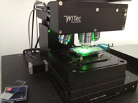
Atomic Force Microscopy coupled with Confocal Raman Microscopy

The Gala platform is equipped to a Atomic Force Microscopy (AFM) coupled with a Confocal Raman Microscopy (RAMAN). Raman is a high-resolution imaging technique that is widely used for the characterization of materials and specimens in terms of their chemical composition.
The AFM-RAMAN (Alpha 300AR microscopy) equipment can giveinformations about the structural and chemical composition of galenic compounds (tablets and pills for example) or other solid systems.
The AFFM technology gives informations about the topography (2D in surface and 3D in volume). Coupled with Raman technology, the microscope gives chemical composition.
This method of characterisation is no-destructive and there is no particular sample preparation. The materials can be analysed at ambiant conditions, under controlled atmosphere or in liquid environment.
Technical specifications:
-
Binocular microscope with a colour camera;
-
3 objectives: 100x (0.9 Numerical Aperture); 50x (0.75 NA); 20x (NA); 10x (0.25 NA);
-
the laser emits an 532 nm wavelength with an adjustable power output to optimise the intensity of the flow on the sample;
-
spectrometer UTHS 300 with a 300 mm focal;
-
Charge Coupled Device camer (CCD)
Raman Mode
-
Acquisition of the full spectrum for each pixel of the image.
-
Confocal microscopy in reflection, confocal imaging in fluorescence.
-
Maximal sample dimension of 120mm length and 25mm width.
-
Spectral wavelength: 442 to 785 nm in excitation, 175 to 4000 cm-1 in detection.
AFM Mode
-
Three modes: contact mode, taping mode and Pulse Force Mode.
-
A Field Programmable Array (FPGA) monitors the whole instrument (CCD camera, Spectrograph, motorised movements of the sample, AFM …) and its extensions.
-
Commun data acquisition software for Raman and AFM modes
